Abnormal uterine bleeding is a common gynaecological problem. The causes of abnormal uterine bleeding are numerous and a comprehensive discussion is beyond the scope of this chapter.
The common causes of uterine bleeding that can be diagnosed and treated by hysteroscopy are:
1) Endometrial polyps
2) Submucous fibroids
3) Endometrial hyperplasia
4) Endometrial cancer
5) Retained products of conceptus
6) Sacculation of a caesarean scar
1) Endometrial polyps
Endometrial polyps are common and may cause abnormal uterine bleeding. Diagnosis of these polyps can be done by transvaginal ultrasound, saline infusion sonography or hysteroscopy (see chapter 6). Polypectomy can be done either during Diagnostic Office Hysteroscopy or hysteroscopy under general anaesthesia (see chapter 37).
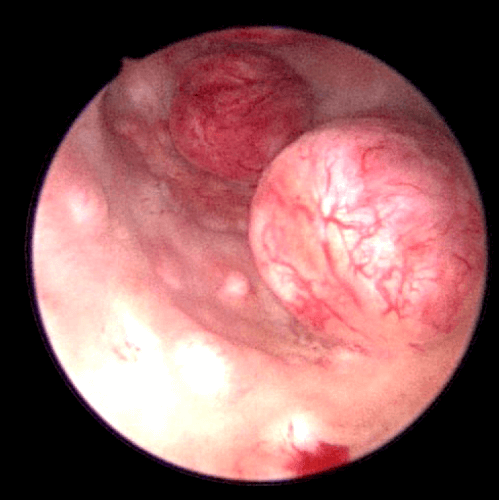
2) Submucous fibroids
Fibroids that protrude into the endometrial cavity are called submucous fibroids Submucous fibroids can be resected hysteroscopically (see chapter 41).
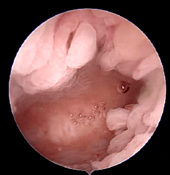
3) Endometrial Hyperplasia
There are many types of endometrial hyperplasia. Some endometrial hyperplasia can progress to endometrial cancer. When the endometrium is thick, especially in post menopausal women, this condition may be suspected. Sometimes, hyperplasia coexists with endometrial cancer. Making a diagnosis of endometrial hyperplasia by hysteroscopy can be difficult. This is because when distension fluid is used during hysteroscopy, the endometrium is compressed, making the hyperplastic tissues more difficult to visualize. Hyperplasia can be focal or global. Endometrial hyperplasia can be suspected when;
1. Focal or papillary mucosal projections with or without gland cysts are seen
2. Abnormal vascular network with atypical vessels are seen
3. Crowded or abnormally spaced gland openings are seen
Taking a biopsy of the abnormal areas and doing a curettage of the endometrium will confirm the diagnosis of endometrial hyperplasia.
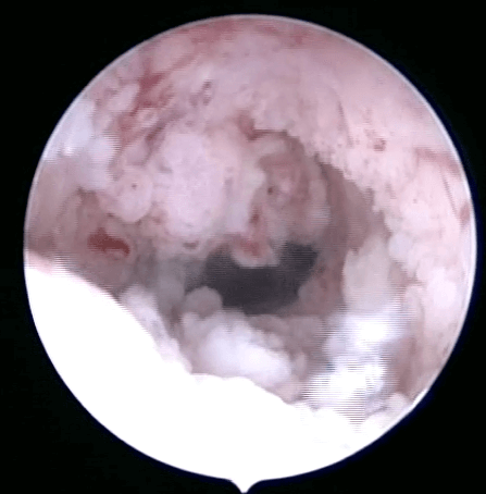
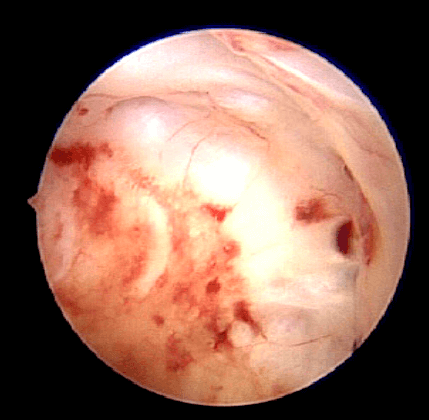
4) Endometrial cancer
The diagnosis of endometrial cancer requires histology. During hysteroscopy, endometrial cancer can be suspected when:
1. Papillary, polypoidal, nodular or mixed endometrial growth is seen with friable tissue
2. There is a focal necrosis of tissues 3. Atypical vessels can be seen
Biopsies need to be taken with an office hysteroscope, the amount of tissue obtained using a fine 5 French biopsy grasper is small. Traditionally, curettage is done to obtain sufficient amount of tissue so as to be more accurate and to obtain
a bigger and deeper amount of endometrial tissue.
5) Retained Products of Conceptus
Retained products of conceptus must always be considered when irregular bleeding is seen in a woman of reproductive age. This usually occurs close to a time of a recent pregnancy but may occur months and even years later. Retained pieces of fetal bone may be seen on ultrasound as an intensely bright line within the endometrium. A hysteroscopy sometimes aided by a wire loop can dislodge these bony fragments.
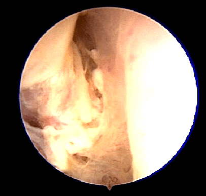
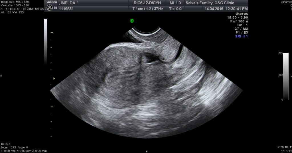
5) Sacculations of a Caesarean section scar
Some women have irregular menses with vaginal spotting or discharge after menstruation. These women sometimes experience fluid collection at the area of the caesarean section scar. This sacculation (g) can be viewed on an ultrasonogram. Hysteroscopy can be performed to look at the caesarean section scar to see whether there is any defect or endometrial tissue adherent to the scar. This is usually a benign condition that may not require any treatment but if persistent, repair of the sacculation can be done by laparoscopy or laparotomy.
Summary
Abnormal uterine bleeding is a common gynaecological problem. Hysteroscopy assists in evaluating the uterine cavity for pathology. In some patients this pathology can be treated by hysteroscopy.