Basic Anatomy
In order to understand gynaecological diseases and gynaecological laparoscopic surgery, it is important to first understand the normal female pelvic anatomy.
The female reproductive system consists of 4 major parts: the uterus, vagina, fallopian tubes and ovaries.
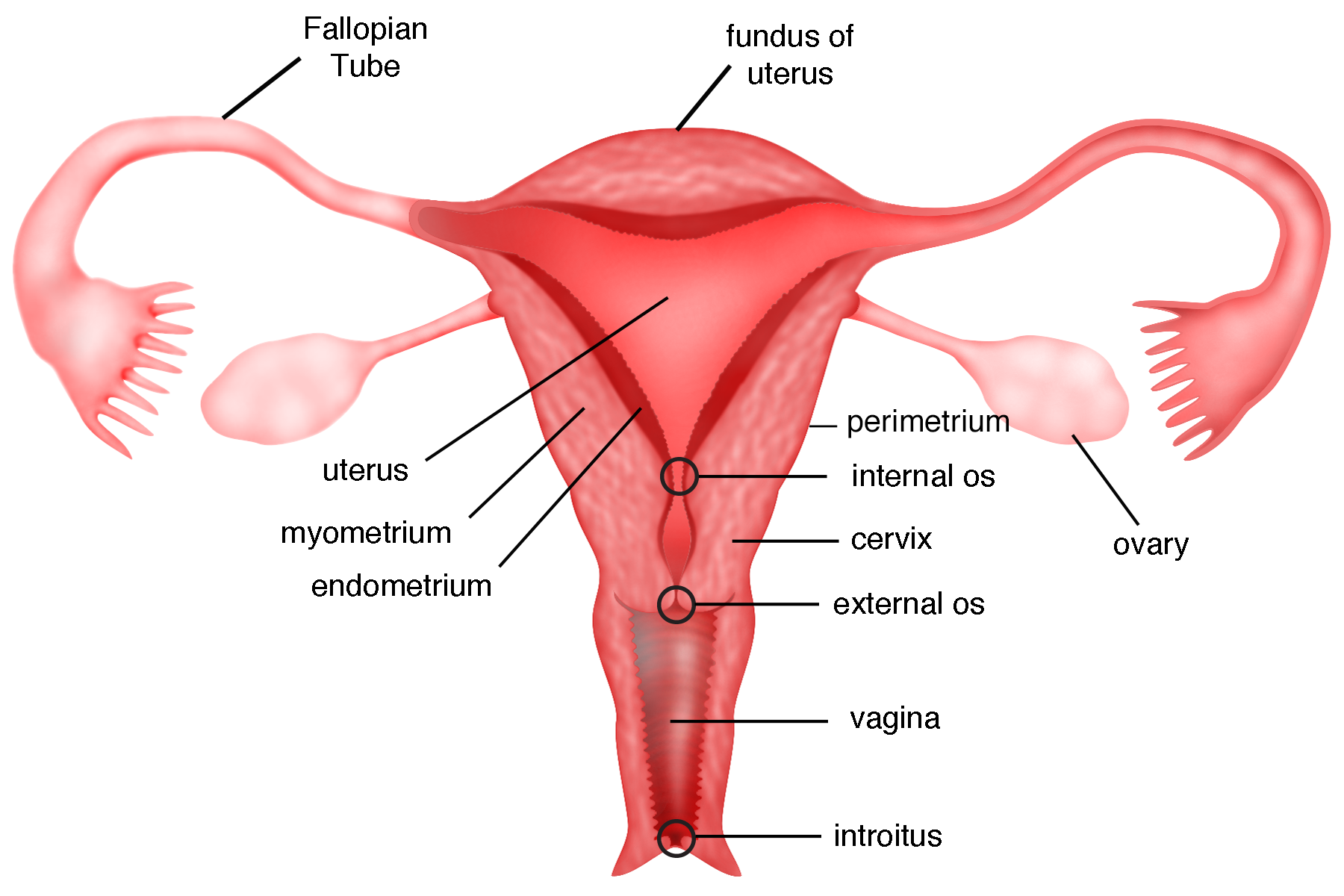
Uterus The uterus, or womb, is a hollow, pear-shaped organ with a thick muscular wall. It is subdivided into two parts: the corpus (body), and the cervix (neck).
The corpus comprises the fundus, which is the top portion of the uterus; and the cavity of the uterus. The cavity is where the embryo/foetus develops during pregnancy. The inner layer or the lining of the uterus, is called the endometrium. Every month, it thickens in preparation for potential pregnancy and sheds during menstruation if pregnancy does not occur. The middle layer of the uterus is known as the myometrium. It is mainly composed of smooth muscle cells, which collectively give the uterus the strength to contract and expel the foetus during childbirth. The outermost layer of the uterus is the serosa, also known as the perimetrium. The cervix is the lower constricted segment of the uterus that joins the upper part of vagina. The small cervical opening into the vagina is called the external os while the one in the uterine cavity is called the internal os. They allow the sperm to enter the uterus during sexual intercourse and the menstrual fluid to flow out of the uterus during menstruation. The cervix can be visualised from the vagina.
- Vagina The vagina is a muscular, narrow canal that extends from the vaginal opening called introitus, to the cervix. It is also known as the birth canal due of the fact that the foetus passes through it to be born during natural childbirth. The inner wall of the vagina is surfaced with numerous folds of soft elastic mucous membrane called vaginal rugae. This allows the vagina to expand considerably during sexual intercourse or childbirth. During menstruation, the vagina provides a channel for the menstrual fluid to flow out of the body.
- Ovaries The ovaries are small, oval-shaped paired glands that are attached to each side of the uterus via a thin, fibrous ovarian ligament. The pair is responsible for storing and nurturing immature egg cells to become mature eggs. Every month, one of the ovaries releases a mature egg into its neighbouring Fallopian tube. In addition to producing eggs, the ovaries produce two main female sex hormones: the oestrogen and progesterone, which are vital in regulating menstrual cycles.
- Fallopian tubes The fallopian tubes, sometimes simply called tubes, are the two channels that connect the ovaries to the uterus. They are the main structures that facilitate fertilization. Each tube is divided into 5 main portions:
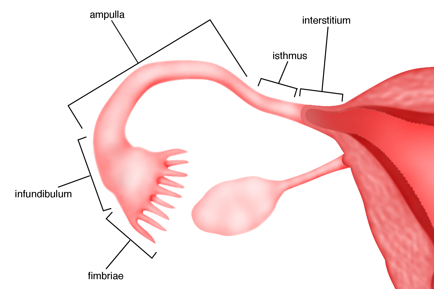
- Fimbriae: The fringe-like structure located at the end of the tube that captures an egg released from the ovary and draws it into the tube.
- Infundibulum: The funnel-like structure of the tube, which is margined by the fimbriae.
- Ampulla: The longest portion of the tube with a thin wall (almost muscle-free) and wide lumen (g). It is usually the portion where fertilization takes place.
- Isthmus: The almost straight portion of the tube with a relatively thick muscular wall and with the narrowest lumen(g).
- Interstitium: The portion of the tube that is closest to the uterus. It is sometimes known as the uterine portion of the tube for the fact that it lies within the uterus. The inner lining of the fallopian tube is made up of fine finger like projections called the cilia. These cilia are important in assisting the movement of the eggs towards the uterine cavity and the sperms into the ampulla of the fallopian tube.
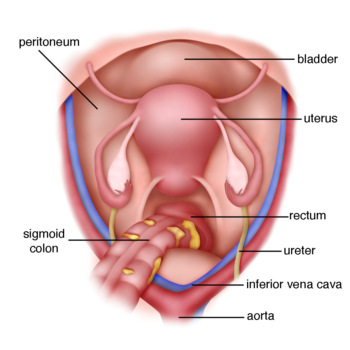
Figure 1.4 is a different view of the female pelvis. It is how the pelvis looks when
a gynaecologist looks at the pelvis either through a laparotomy (large incision on the abdomen) or through a laparoscope (key hole surgery).
The uterus is in the centre. In front (anterior) of the uterus is the urinary bladder. Behind (posterior) to the uterus is the rectum. A slippery membrane called the peritoneum(g) covers the whole pelvis and abdomen. Beneath the peritoneum, on either side of the pelvis, run the ureters. The ureter is a small tube that carries urine and runs from the kidney to the bladder. Large blood vessels are present on both sides of the pelvis. These blood vessels carry blood from the heart to both the legs and back.
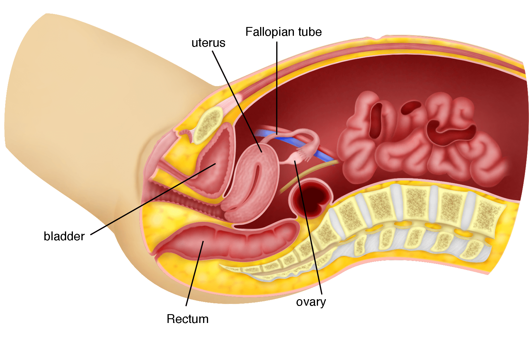
Figure 1.5 is a side view of the abdomen and pelvis. The uterus is in the centre. On the side and behind the uterus are the fallopian tubes and the ovaries. In front of (anterior to) the uterus is the urinary bladder and behind it (posterior to) is the rectum.
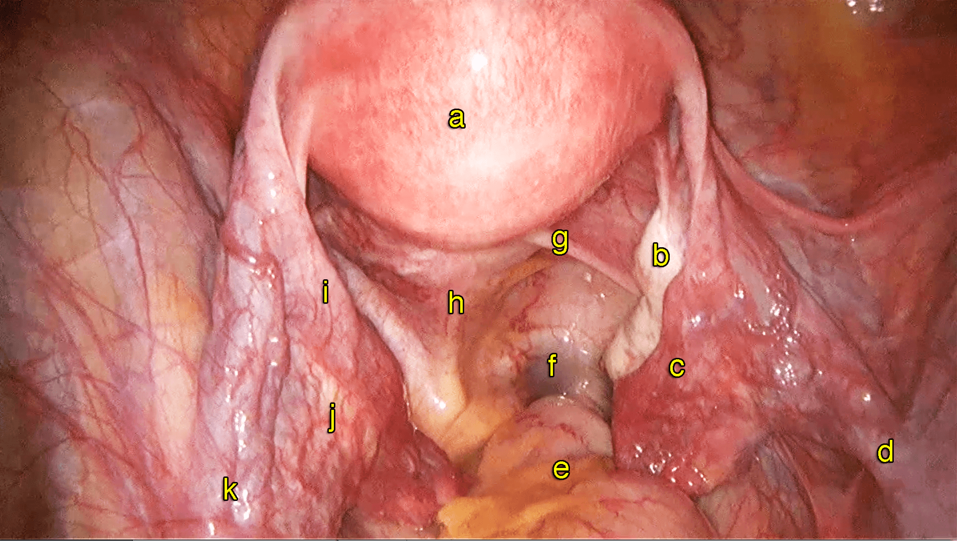
(a) Uterus,
(b) Right ovary,
(c) Right fallopian tube,
(d) Right infundibulopelvic ligament,
(e) Rectum,
(f) Pouch of Douglas,
(g) Right uterosacral ligament,
(h) Left uterosacral ligament,
(i) Left fallopian tube,
(j) Left ovary,
(k) Leftinfundibulopelvicligament.
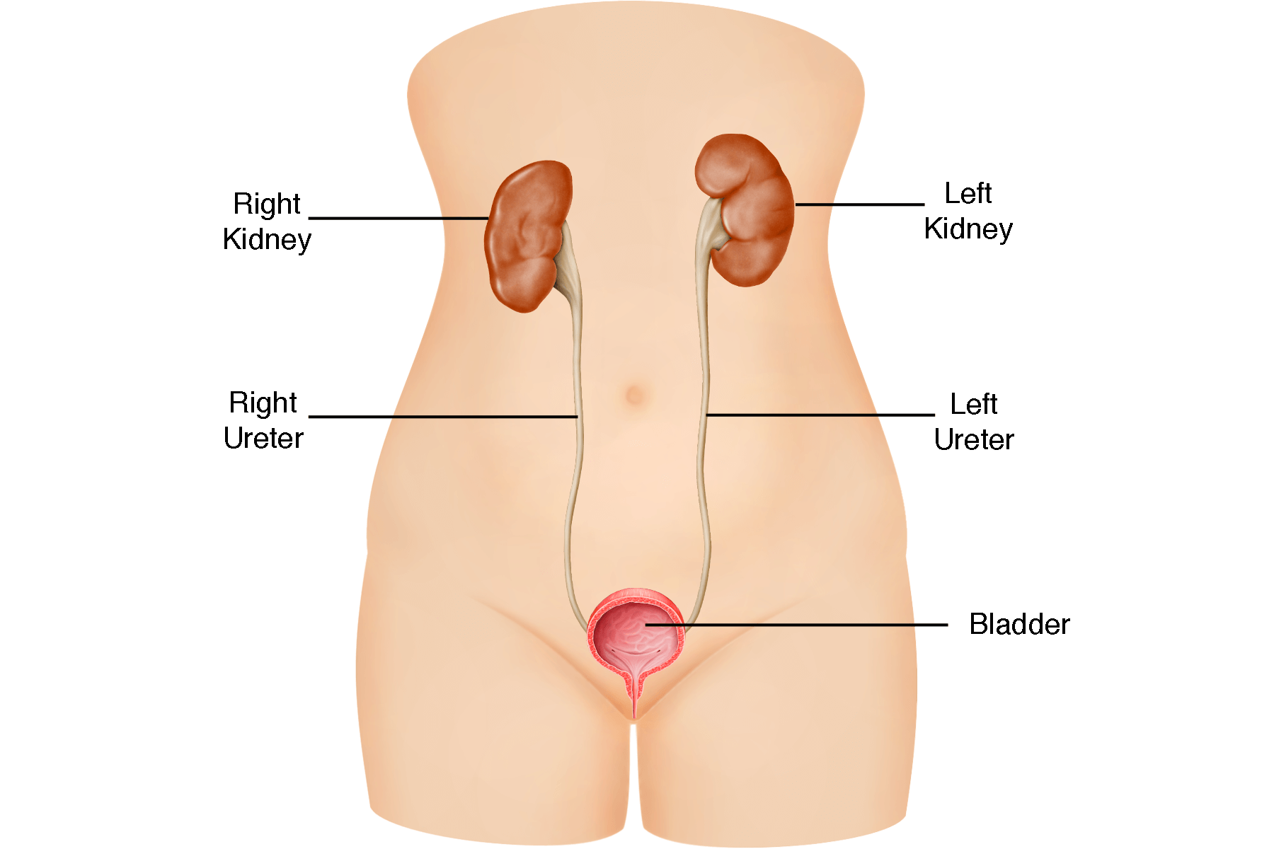
Figure 1.7 shows the urinary system. Ureters connect the kidneys to the urinary bladder. Ureters are found behind the peritoneum on the pelvic sidewalls. They run below the ovaries and on the side of the cervix under the uterine arteries before entering the bladder via the ureteric tunnel. Ureters are important structures to identify during gynaecological surgeries.

Normal female pelvic anatomy.
https://vimeo.com/149588511
https://www.youtube.com/watch?v=i4YpzdhydpQ&t=4s
Summary
It is important to understand the normal female anatomy before proceeding to learn about common gynaecological diseases and laparoscopic surgery for gynaecological diseases.